

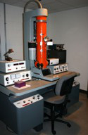
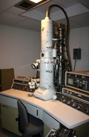
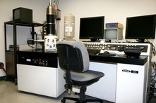
The Department of Biology at the University of Maryland, College Park maintains the Laboratory for Biological Ultrastructure, a high-quality, well-equipped biological imaging facility. For more than 30 years, this facility has been an important resource for university faculty and students. The multi-room supervised facility contains two transmission (Zeiss EM 10 CA and JEOL 100CX II) and two scanning (Hitachi S-4700 and Amray 1820D) electron microscopes. Light microscopic instrumentation includes two Zeiss photomicroscopes equipped with bright field, phase contrast, differential interference contrast and polarized light optics. A recently acquired Bio-Rad MRC 1024 confocal microscope has been added to provide fluorescent and laser scanning capabilities. Although the laboratory is housed in and funded by the College of Computer, Mathematical, & Natural Sciences, it is open to use by other researchers of the university community. Investigators from the following departments benefit from the facility's state-of-the-art equipment:
- Animal and Avian Sciences
- Biology
- Cell Biology and Molecular Genetics
- Chemistry and Biochemistry
- Chemical Engineering
For more information:
Laboratory for Biological Ultrastructure
Timothy Maugel, Director
0240 Biology-Psychology Building
tmaugel@umd.edu
Phone: 301.405.6898
Fax: 301.314.9358
Equipped with a tilt stage and Dage 68 high-resolution analog video camera.
Equipped with a scanning attachment.
Equipped with a EDAX Genesis energy dispersive x-ray microanalysis
system, Robinson backcattered electron detecto, Robinson Chamber
View camera system, Pic I digital image aquisition system and a
Seikosha video printer.
Equipped with Hitachi cryo-stage, Centaurus backscattered electron
detector and GW chamberscope camera.
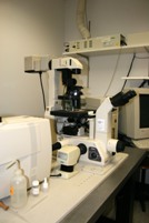
Diaphot 300 inverted microscope
Equipped with DIC and fluorescent optics.
Two Zeiss Photomicroscope II
Equipped with bright field, Normarski differential interference, phase contrast
and polarizing optics.
Two Zeiss RA compound microscopes and five Wild and Bausch & Lomb
dissecting microscopes
osmometer, shakers, dissecting and compound microscopes, ect., is maintained in operating condition within the Laboratory.


Light microscopic images can be recorded in film(35mm or sheet film), video or digital formats. High resolution flatbed and slide scanners are available for converting analog images to digital format. Video, laser, inkjet, thermal and dye-sub printers are available to provide photographic prints of images from LM, SEM or TEM. Complete wet photographic facilities, including film and plate processing, print production on a point source enlarger, and trimming and mounting equipment are contained within the facility for the preparation of publication ready material.